Education
Travel
New transmission electron microscope arrives at Montana Tech
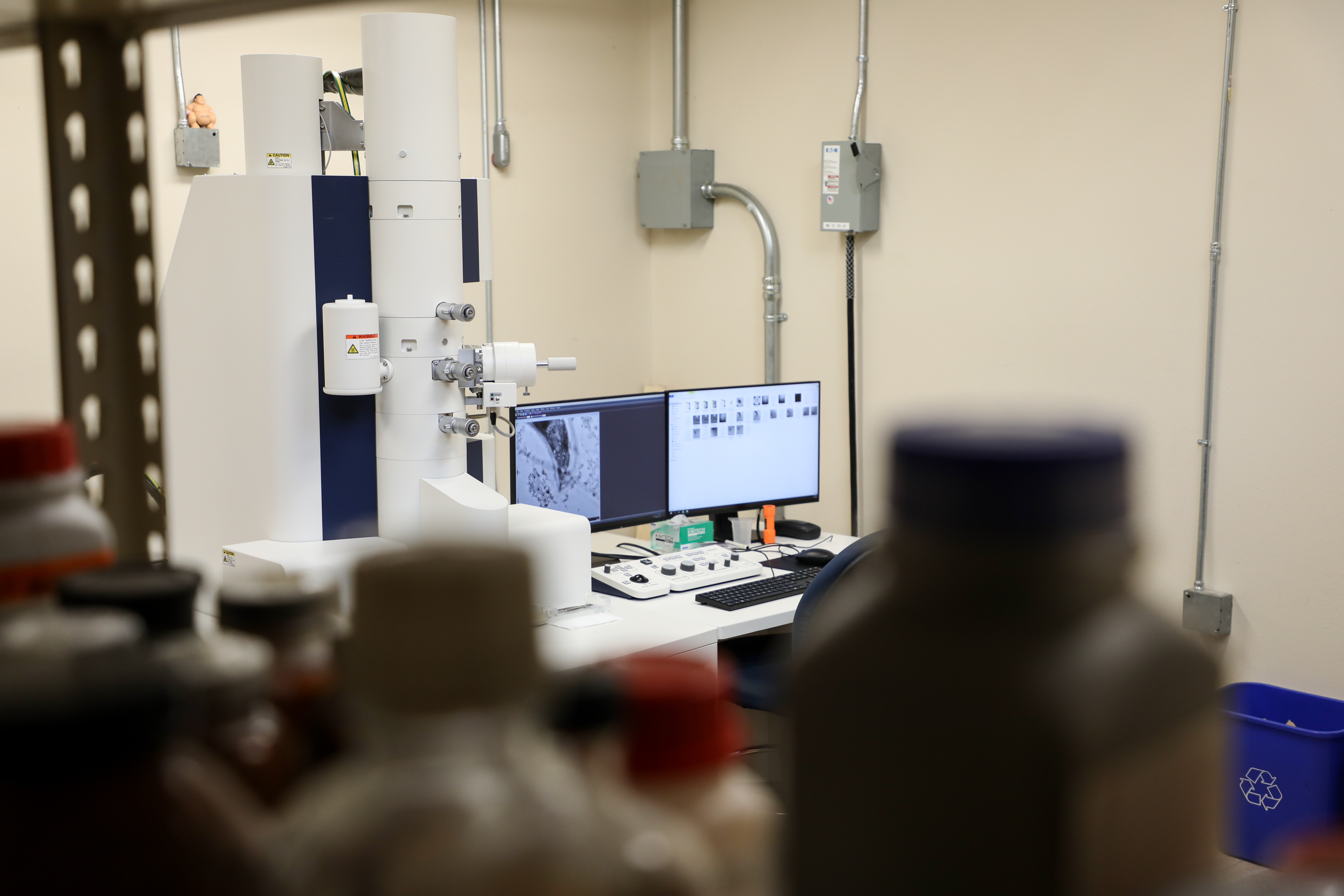
Researchers at Montana Technological University will be able to better explore worlds invisible to the naked eye after the delivery of a multifunctional transmission electron microscope. The new equipment will significantly expand opportunities for the campus community, state of Montana, and Rocky Mountain West.
Montana Tech purchased the $1 million microscope with funds from the National Science Foundation that were acquired via a highly competitive grant process. On September 25, Montana high school teachers will experience the new microscope. The media is invited to attend.
The Hitachi HT7820 Transmission Electron Microscope (TEM) is the latest model of its kind, which means that it is among the best in its class, and a huge win for scientists in the region.
“This instrument has capabilities that other instruments in the state and region don’t have,” Dr. Marissa Pedulla, the primary grant author, said.
Pedulla has a collection of images of bacteriophages, which are viruses that infect and replicate in bacterial cells. Many of the images were created at the University of Montana, located 120 miles away, where Pedulla and her students formerly traveled to in order to conduct microscopy needed for her work previously. Dr. James Driver was the microscopist who worked with Dr. Pedulla at UM, but he has joined the Montana Tech staff as the microscopist at the helm of the new instrument. Grant funding will keep Driver on staff for at least 26 months.
Driver, who worked for UM for over two decades, speaks of the new microscope in a tone of admiration. “This instrument is fantastic. It has state-of-the-art imaging of viruses and the inside of cells and components that allow us to see surface structures on a sample similar to a scanning electron microscope (SEM) as well as the elements within that sample,” said Driver. “At the elemental level, it can tell us if there is lead or arsenic inside a cell.”
The tool is so powerful it can target individual proteins in subcellular structures. The new capability is expected to be a boon to numerous departments at Montana Tech, ranging from materials science to biology, chemistry and environmental engineering. Seeing materials or phages with such detail is important because researchers can understand more about their structure, chemical nature and mechanisms of synthesis and proliferation.
Pedulla is excited to share the microscope’s capabilities with students in her PHAGES program. The program has allowed thousands of Montana school children to explore the world of bacteriophages. The program is funded by a National Institutes of Health grant. More than 100 students have discovered previously unknown bacteriophages in Pedulla’s lab, classes, and in outreach visits to Montana K-12 classrooms. The new microscope will allow students to have even more capacity to probe into the microscopic universe.
“I would just like to thank all of the students and teachers who have participated in phage discovery over the past 18 years,” Pedulla said. “They’ll be able to see their discoveries through this microscope. I would also like to thank my co-investigators on the grant.”
Pedulla’s grant co-authors are Drs. Katie Hailer, Jack Skinner, and Jessica Andriolo. The researchers say the microscope is expected to serve the community for decades.
“There is just so much that can be done with this modern scientific instrument,” Driver said.