E.M.Tech - Electron Microscopy Laboratory- Image Gallery

Gold Particles on Holey Carbon Grid
Hitachi HT 7820 Scanning Transmission Electron Microscope Image. Size bar 500 nm.

Tooj Phage
Hitachi HT7820 TEM image. MT Tech NIH-SEPA Phages Program. Size bar 500 nm.

Daggertail Phage
Hitachi HT7820 TEM image. MT Tech NIH-SEPA Phages Program. Size bar 200 nm.

Duckfeet Phage - Family Myoviridae
Hitachi HT7820 TEM image. MT Tech NIH-SEPA Phages Program. Size bar 100 nm.

Daka Phage
Hitachi HT7820 TEM image. MT Tech NIH-SEPA Phages Program. Size bar 200 nm.
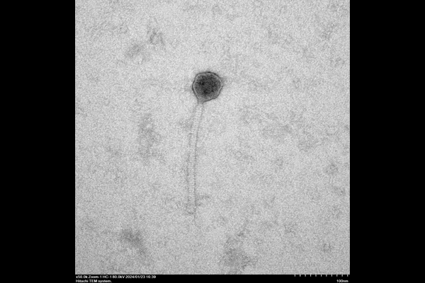
Novi-14 Phage
Hitachi HT7820 TEM image. MT Tech NIH-SEPA Phages Program. Size bar 100 nm.
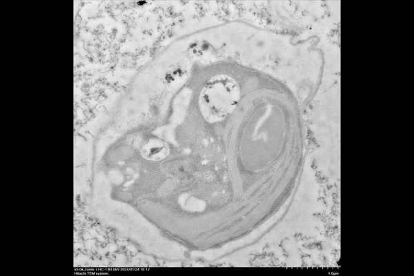
Algal Cell with Carbon Nanotubes conjugated with AgNO3
Hitachi HT7820 TEM image. Size bar 1 um.

Algal Cell with Carbon Nanotubes conjugated with AgNO3
Hitachi HT7820 TEM image. Size bar 500 nm.
Connect with E.M.Tech.
To learn more, or to access or schedule time on the HT7820 contact us today.
Dr. Jim Driver
Electron Microscopist
EMsignup@mtech.edu
Electron Microscopist
EMsignup@mtech.edu